Lower Body Bone Diagram : Bones Of The Lower Limb Anatomy And Physiology I / Teachme anatomy part of the teachme series the medical information on this site is provided as an information resource only, and is not to be used or relied on for any diagnostic or treatment purposes.
byAdmin-
0
Lower Body Bone Diagram : Bones Of The Lower Limb Anatomy And Physiology I / Teachme anatomy part of the teachme series the medical information on this site is provided as an information resource only, and is not to be used or relied on for any diagnostic or treatment purposes.. Femur (2) tibia (2) fibula (2) patella (2) tarsals (14) metatarsals (10) phalanges (28) total number of bones=60. The knee joint is the largest joint in the body and is primarily a hinge joint, although some sliding and rotation occur. Key bones in the abdominal area include the base of the ribcage and the lumbar spine in the lower back. The tibia (shin bone) is the medial bone of the leg and is larger than the fibula, with which it is paired (figure 6.52). The sternum, commonly known as the breastbone, is a long, narrow flat bone that serves as the keystone of the rib cage and stabilizes the thoracic skeleton.
The back supports the weight of the body, allowing for flexible movement while protecting vital organs and nerve structures. Your lower back (lumbar spine) is the anatomic region between your lowest rib and the upper part of the buttock. The bones of the legs are those that make up the thigh, the lower half of the legs, and the feet. Check spelling or type a new query. Related posts of anatomy of lower body anatomy of males/females.
Lower Limb Bones Illustrations Radiology Case Radiopaedia Org from prod-images-static.radiopaedia.org Posteriorly the body is connected to a thin ring of bone known as the arch. Key bones in the abdominal area include the base of the ribcage and the lumbar spine in the lower back. Maybe you would like to learn more about one. There are two hip bones, one on the left side of the body and the other on the right. The long bones of the body contain many distinct regions due to the way in which they develop. The lower leg contains two major long bones, the tibia and the fibula, which are both very strong skeletal structures. The tibia (also called the shinbone) is located near the midline of the leg. Balance the weight of your head on top of your spine.
There are two hip bones, one on the left side of the body and the other on the right.
Female anatomy includes the external genitals, or the vulva, and the internal reproductive organs. The long bones of the body contain many distinct regions due to the way in which they develop. The lower leg contains two major long bones, the tibia and the fibula, which are both very strong skeletal structures. Bone diagram forehead (frontal bone) nose bones (nasals) cheek bone (zygoma) upper jaw (maxilla) lower jaw (mandible) breast bone (sternum) upper arm bone (humerus) lower arm bone (ulna) thigh bone (femur) collar bone (clavicle) toe bones (phalanges) ankle bones. Together, they form the part of the pelvis called the pelvic girdle. The muscles of the lower back help stabilize, rotate, flex, and extend the spinal column, which is a bony tower of 24 vertebrae that gives the body structure and houses the spinal cord. There are two hip bones, one on the left side of the body and the other on the right. 1 your spine in this region has a natural inward curve. The knee joint is the largest joint in the body and is primarily a hinge joint, although some sliding and rotation occur. The bones of the legs are those that make up the thigh, the lower half of the legs, and the feet. Related posts of muscles of the lower back and buttocks diagram abdominal muscle diagram. We did not find results for: A cylinder of bone known as the vertebral body makes up the majority of the lumbar vertebrae's mass and bears most of the body's weight.
The knee joint is the largest joint in the body and is primarily a hinge joint, although some sliding and rotation occur. Maybe you would like to learn more about one. In all, there are believed to be 80 organs in your body, all serving different functions and uses. Check spelling or type a new query. The bones of the pelvis and lower back work together to support the body's weight, anchor the abdominal and hip muscles, and protect the delicate vital organs of the vertebral and abdominopelvic cavities.
Skeletal System Anatomy And Physiology Nurseslabs from nurseslabs.com In all, there are believed to be 80 organs in your body, all serving different functions and uses. Several muscles that move the arms, head, and neck have their origins on the sternum. 1 your spine in this region has a natural inward curve. We did not find results for: Female anatomy includes the external genitals, or the vulva, and the internal reproductive organs. The vertebral column of the lower back includes the five lumbar vertebrae, the sacrum, and the coccyx. The sternum, commonly known as the breastbone, is a long, narrow flat bone that serves as the keystone of the rib cage and stabilizes the thoracic skeleton. Anatomy of males/females 12 photos of the anatomy of males/females anatomy of male and female, anatomy of male and female brain, anatomy of male and female breasts, anatomy of male and female reproductive organs, explanation of anatomy and reproduction in males and females, human anatomy, anatomy of male and female, anatomy.
Evenly distribute weights from your upper body into the lower extremities.
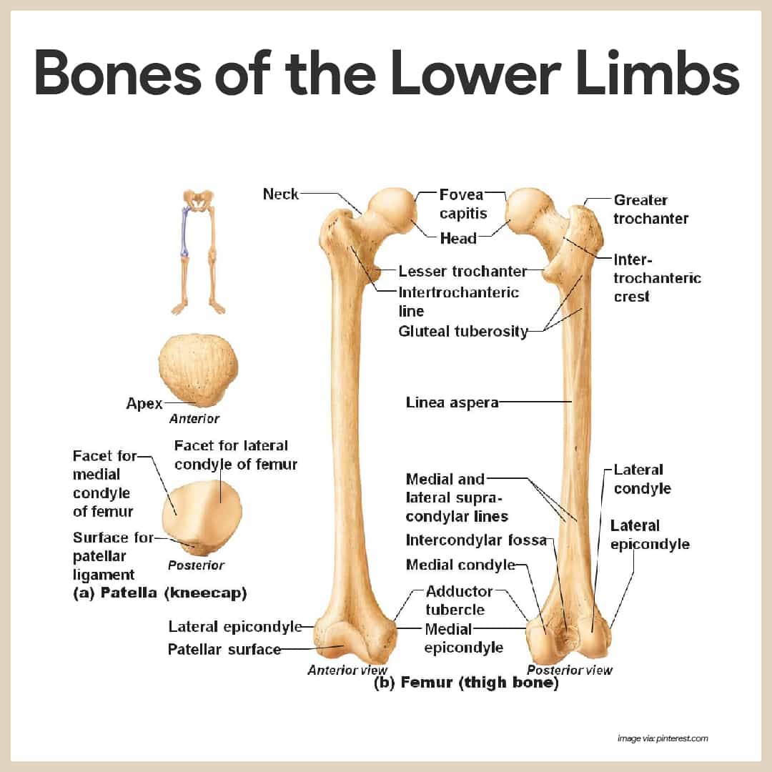
This curve, called lordosis, helps to: They hold up your body, and along with your muscles, keep you moving. The arch surrounds the hollow vertebral foramen and connects the body to the bony processes on the posterior of the vertebra. Your body organs range from your brain, heart, liver, skin, lungs, kidneys, intestines, stomach, bladder, etc. Abdominal muscle diagram 12 photos of the abdominal muscle diagram abdominal muscle anatomy bodybuilding, abdominal muscle diagram female, abdominal muscle groups diagram, human abdominal muscle diagram, lower abdominal muscle diagram, human muscles, abdominal muscle anatomy bodybuilding, abdominal. Balance the weight of your head on top of your spine. Related posts of anatomy of lower body anatomy of males/females. Related posts of muscles of the lower back and hip diagram body muscles with names. The muscles of the lower back help stabilize, rotate, flex, and extend the spinal column, which is a bony tower of 24 vertebrae that gives the body structure and houses the spinal cord. Body muscles with names 12 photos of the body muscles with names body muscles and names, body muscles and their names, body muscles parts name, human body muscles with names, muscular body parts name, human muscles, body muscles and names, body muscles and their names, body muscles parts name, human body. One of the body's largest and longest nerves is the sciatic nerve. The tibia (shin bone) is the medial bone of the leg and is larger than the fibula, with which it is paired (figure 6.52). This is the longest bone in the human body, and is also known as the thigh bone.
This diagram depicts abdominal regions areas lower.human anatomy diagrams show internal organs, cells, systems, conditions, symptoms and sickness information and/or tips for healthy living. This diagram depicts anatomy of the lower leg achilles tendon.human anatomy diagrams show internal organs, cells, systems, conditions, symptoms and sickness information and/or tips for healthy living. Also called the shin bone, the tibia is the longer of the two bones in the. Do you ever wonder what the major organs of the body are and. This curve, called lordosis, helps to:
Cybersurgeons from www.e-missions.net The back supports the weight of the body, allowing for flexible movement while protecting vital organs and nerve structures. Key bones in the abdominal area include the base of the ribcage and the lumbar spine in the lower back. The bones of the legs are those that make up the thigh, the lower half of the legs, and the feet. The muscles of the lower leg can divided into 3 main groups: The knee joint is the largest joint in the body and is primarily a hinge joint, although some sliding and rotation occur. The vertebral column of the lower back includes the five lumbar vertebrae, the sacrum, and the coccyx. Your lower back (lumbar spine) is the anatomic region between your lowest rib and the upper part of the buttock. The long bones of the body contain many distinct regions due to the way in which they develop.
The back supports the weight of the body, allowing for flexible movement while protecting vital organs and nerve structures.
From i.pinimg.com check spelling or type a new query. Your body organs range from your brain, heart, liver, skin, lungs, kidneys, intestines, stomach, bladder, etc. Together, they form the part of the pelvis called the pelvic girdle. The back supports the weight of the body, allowing for flexible movement while protecting vital organs and nerve structures. The tibia (shin bone) is the medial bone of the leg and is larger than the fibula, with which it is paired (figure 6.52). They hold up your body, and along with your muscles, keep you moving. Several muscles that move the arms, head, and neck have their origins on the sternum. Anatomy of males/females 12 photos of the anatomy of males/females anatomy of male and female, anatomy of male and female brain, anatomy of male and female breasts, anatomy of male and female reproductive organs, explanation of anatomy and reproduction in males and females, human anatomy, anatomy of male and female, anatomy. Evenly distribute weights from your upper body into the lower extremities. One of the body's largest and longest nerves is the sciatic nerve. The muscles of the lower leg can divided into 3 main groups: It descends from the sacral plexus through the buttocks and into the thighs to supply nerve impulses to and from the muscles and skin in the hip joints and thighs, the lower legs, feet and most of the skin below the knee. The muscles of the lower back help stabilize, rotate, flex, and extend the spinal column, which is a bony tower of 24 vertebrae that gives the body structure and houses the spinal cord.
Anatomy of males/females 12 photos of the anatomy of males/females anatomy of male and female, anatomy of male and female brain, anatomy of male and female breasts, anatomy of male and female reproductive organs, explanation of anatomy and reproduction in males and females, human anatomy, anatomy of male and female, anatomy lower body diagram. Related posts of muscles of the lower back and buttocks diagram abdominal muscle diagram.